教 AI 理解对称性为何能让产前诊断更可靠
MIT 与波士顿儿童医院的研究人员开发的一款模型可实时调整扫描以追踪胎儿运动,有望让家庭免于反复往返医院。
Alex Ouyang|Abdul Latif Jameel Clinic for Machine Learning in Health
Abdul Latif Jameel Clinic for Machine Learning in Health
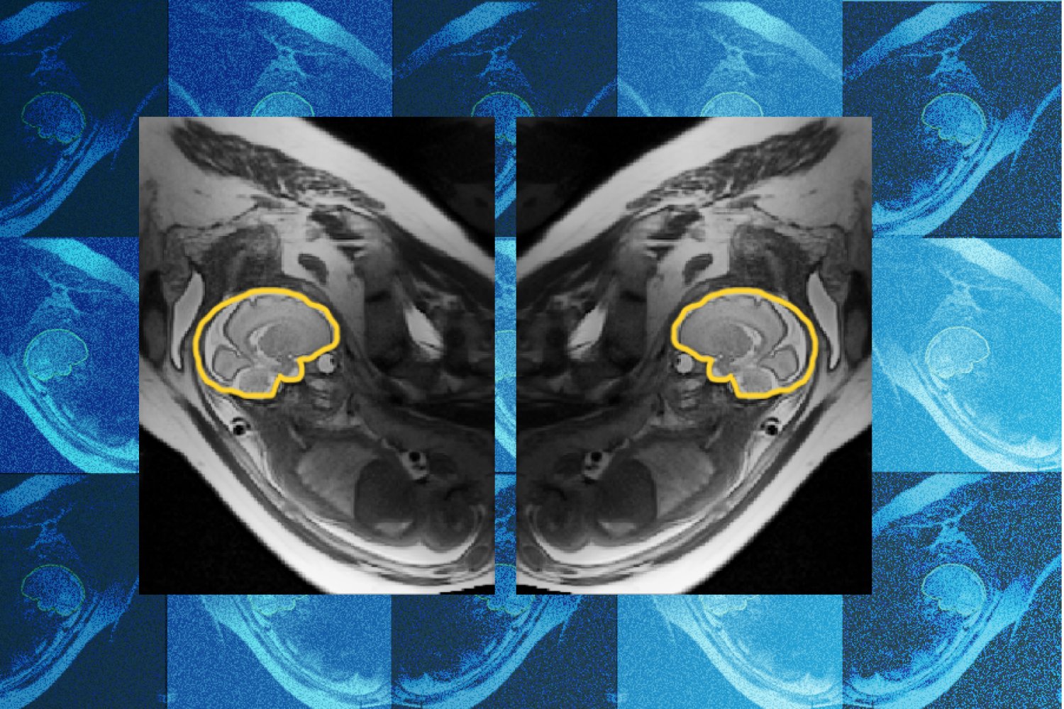
矢状位下胎儿脑部的 MRI 扫描图像(以黄色轮廓标出)。E(3)-Pose 是一款有助于在胎儿脑部 MRI 中追踪运动的模型。该模型可帮助临床医生更早、更准确地诊断发育异常。
图片由 MIT Jameel Clinic 及研究人员提供。
尽管经历了数百万年的进化,人类胎儿的发育仍是一个复杂的过程,容易受到遗传、环境和代谢紊乱的影响。这些紊乱可能导致发育异常,而最先受影响的器官之一,正是胎儿最早开始发育、并在出生后仍继续发育的器官:大脑。
在常规产前超声检查中,超声技师会标注出可能存在的脑部异常,以便进一步进行磁共振成像(MRI)检查。但不够完美的成像可能导致漏诊或误诊。
为帮助这些异常的早期发现与治疗,MIT、波士顿儿童医院和 Inria 的研究人员开发并训练了一款名为 E(3)-Pose 的深度学习模型,可在胎儿头部运动时对其进行成像,从而捕获可供放射科医生查看的高分辨率图像。相关论文已在 2026 年欧洲计算机视觉会议(European Conference on Computer Vision)上发表。
“胎儿移动带来的一个问题在于,切片位置到处都是,”第一作者 Ramya Muthukrishnan 说。Muthukrishnan 是 MIT 电气工程与计算机科学 Sunlin (1966) and Priscilla Chou 讲席教授 Polina Golland 实验室的博士生;Golland 是 MIT Abdul Latif Jameel Clinic for Machine Learning in Health(Jameel Clinic)和 MIT 计算机科学与人工智能实验室(CSAIL)的首席研究员。
Muthukrishnan 所说的切片,指的是胎儿 MRI 扫描过程中拍摄的一系列 2D 图像。这些 2D 平面叠加在一起,实际上构成了胎儿头部的单幅高分辨率 3D 图像。但如果头部在成像过程中发生移动,每张 2D 图像“切片”可能最终是从不同角度拍摄的,导致最终扫描出现缺口以及切片方向异常。这些问题可能导致审阅 MRI 的医生漏诊或误诊胎儿脑部病变。
“有些非常细微的细节,需要一幅非常好的脑部立体图像才能看到,”Muthukrishnan 解释道。“如果有覆盖缺口,你就看不到关键区域,而你需要看得清楚,才能发现发育异常。”
E(3)-Pose 之所以特别擅长追踪胎儿运动,在于它对物理的重视。模型并非只是通过数据训练来学习物理规律,物理原理本身被嵌入了神经网络:胎儿头部的左右对称性被内嵌于模型架构之中,使模型更容易准确识别脑部的方位。
Muthukrishnan 在博士生涯初期开始参与这个项目时,她没有任何物理学背景,更不用说 MRI 物理学了。她的经验大多在计算机科学领域。“当我不得不去把这个东西部署到扫描仪上时,我很意外,”她回忆道。“我完全没料到我会让扫描仪崩溃这么多次,因为把东西放到硬件上、让它能在硬件上跑起来,和在 GPU 上训练模型处理数据完全是两码事。”
尽管困难重重,Muthukrishnan 还是坚持了下来。她记得在博士初期跟随一位在波士顿儿童医院做胎儿 MRI 扫描的放射科医生时,观察到患者和放射科医生共同的挫败感:怀孕的母亲在 MRI 里待了一个小时后感到不适,结果扫描做到一半就放弃了;放射科医生则无法获得完整、高质量的 MRI 来监测胎儿的发育。
“看到人们为此感到挫败,帮助我决定了自己博士期间想做什么,因为感觉这是一个痛点,”Muthukrishnan 说。
幸运的是,她得到了帮助:共同作者 Borjan Gagoski 是 Harvard Medical School 放射学助理教授、Boston Children's Hospital 研究员,他开发了一套采集额外 MRI 扫描的序列,用于辅助训练 E(3)-Pose,并与 Muthukrishnan 合作,在扫描仪每次崩溃时帮忙排查故障。共同作者、Boston Children's 研究助理 Aryn Lee 招募了这些怀孕志愿者,并协助完成每一次 MRI 扫描的采集。指导该项目的高级作者包括 MIT 电气工程与计算机科学教授 Elfar Adalsteinsson;Harvard Medical School 儿科学 Käthe Beutler 讲席教授兼放射学教授 Ellen Grant;以及 Golland。Inria 研究员、Golland 课题组前博士后 Benjamin Billot 帮助 Muthukrishnan 上手了他最初开发的方法。
“Ramya 的工作将等变性、对称性等几何概念与深度学习的能力优雅地结合起来,改变了 MRI 扫描仪采集图像的方式,最终帮助医生在发育早期发现并处理问题。这是一个令人印象深刻的研究项目,它在图像采集建模方面既深入又基础,同时具有明确的临床影响,”Golland 说。
Grant 是 Boston Children's 胎儿与新生儿神经影像及发育科学中心(FNNDSC)主任,她补充说,Muthukrishnan 的工作“将实现胎儿脑部 MRI 的自动化,以更快、更舒适的扫描取代当今缓慢的人工流程,同时为放射科医生提供质量更高的图像用于判读。”
E(3)-Pose 的应用可能不止于胎儿 MRI 的运动追踪。它还能协助对其他难以成像的解剖结构进行成像,甚至可用于自动驾驶场景——在这些场景中,有限的传感器需要采集含噪声的三维数据,以追踪三维物体。
目前,Muthukrishnan 希望改进 E(3)-Pose,使其能够帮助对孕中期或更早期的胎儿进行成像,因为该模型主要是在孕晚期胎儿的数据上训练的。孕中期的胎儿“非常小,而且因为子宫内空间更大,他们的活动幅度要大得多,”Muthukrishnan 说。“更值得重点关注的是他们,因为脑部异常发现得越早,干预就越容易。”
这项工作得到了 MIT Jameel Clinic、MIT Takeda Program、Chou Family Transformative Research Fund、美国国家生物医学影像与生物工程研究所以及 Eunice Kennedy Shriver 国家儿童健康与人类发展研究所的支持。
通过探究细菌如何劫持细胞功能,副教授 Rebecca Lamason 希望进一步了解宿主细胞是如何执行这些功能的。
一种新方法接续爱因斯坦未竟的工作,用于预测粒子在极端条件下如何在流体中运动。
一个新系统能够识别并纠正机器人记忆中不可靠的物体描述,从而提升机器人与环境交互的能力。
为了缅怀这位长期任教的教授,实验室成员、校友、同事以及其他关心他的人们与他的家人齐聚 MIT 46 号楼,并在线上同步参加。
该团队从 ARPA-H 获得最高 3800 万美元的资助,目标是打造一种低功耗、非侵入式的传感器贴片,可实时测量心力衰竭和脓毒症的生物标志物。
由学生自主运营的 MIT Combat Robotics Club 成员,通过“破坏”来学习。
译文由英文原文机器翻译,术语按行业惯例统一,可在文末展开原文核对。
查看英文原文(Why teaching AI about symmetry could make prenatal diagnosis more reliable)
Why teaching AI about symmetry could make prenatal diagnosis more reliable | MIT News | Massachusetts Institute of Technology
Why teaching AI about symmetry could make prenatal diagnosis more reliable
A model developed by researchers at MIT and Boston Children's Hospital adjusts scans on the fly to track fetal movement, potentially sparing families repeat hospital visits.
Alex Ouyang|Abdul Latif Jameel Clinic for Machine Learning in Health
Abdul Latif Jameel Clinic for Machine Learning in Health

An MRI scan of a fetal brain (outlined in yellow) in the sagittal position. E(3)-Pose is a model that helps track movement in an MRI of the fetus’s brain. The model could help clinicians make earlier and more accurate diagnoses of developmental anomalies.
Image courtesy of the MIT Jameel Clinic and the researchers.
Despite millions of years of evolution, the development of the human fetus remains a complex process that is vulnerable to genetic, environmental, and metabolic disruptions. These disruptions could result in developmental abnormalities, starting with one of the first organs to begin developing in the fetus that continues to develop after birth: the brain.
In a routine prenatal ultrasound, the sonographer will flag possible brain abnormalities for additional magnetic resonance imaging (MRI). But imperfect imaging can result in missed or incorrect diagnoses.
To assist with the early detection and treatment of these abnormalities, researchers at MIT, Boston Children’s Hospital, and Inria have developed and trained a deep learning model called E(3)-Pose that helps image the fetus’s head while it’s in motion to capture a high-resolution image that the radiologist can examine. A paper about the work was presented at the 2026 European Conference on Computer Vision.
“One of the problems with having the baby move is that the slices are all over the place,” says first author Ramya Muthukrishnan. Muthukrishnan is an MIT PhD student in the lab of Sunlin (1966) and Priscilla Chou Professor of Electrical Engineering and Computer Science Polina Golland, who is a principal investigator at the MIT Abdul Latif Jameel Clinic for Machine Learning in Health (Jameel Clinic) and the MIT Computer Science and Artificial Intelligence Laboratory (CSAIL).
By slices, Muthukrishnan is referring to a series of 2D images taken during a fetal MRI scan. These 2D planes stacked together effectively make up one high-resolution, 3D image of the fetus’s head. But if the head shifts during imaging, each 2D image “slice” could end up being taken from a different angle, resulting in gaps in the final scan and unusual slice orientation. These problems may cause the physician reviewing the MRI to miss or incorrectly diagnose a pathology in the fetal brain.
“There are very fine details that you need a very good volumetric picture of the brain to see,” explains Muthukrishnan. “If you have coverage gaps, you can’t see critical regions, and you need to be able to see well to detect abnormal development.”
What makes E(3)-Pose particularly good at tracking fetal motion is its focus on physics. Rather than simply training the model to learn physics from the data, the very principles of physics are baked into the neural network: left-right symmetry of the fetal head is embedded in the model architecture, making it easier for the model to accurately identify the brain’s orientation.
When Muthukrishnan began working on the project at the start of her PhD, she had no background in physics, let alone MRI physics. Most of her experience was in computer science. “I was surprised when I had to go and implement this on the scanner,” she recalls. “I was not prepared for how many times I would crash the scanner because putting things on hardware and getting it to work on hardware is a different beast than training a model to run on data on a GPU.”
Despite the challenges, Muthukrishnan persisted. She remembers shadowing a radiologist who performed fetal MRI scans at Boston Children’s Hospital at the start of her PhD and observing the shared frustration of the patient and radiologist: the pregnant mother felt uncomfortable after spending an hour in an MRI, only to quit the scan halfway; the radiologist was unable to procure a full, high-quality MRI to monitor the development of the fetus.
“Seeing frustration about it helped me decide what I wanted to do with my PhD, because it felt like this was a pain point,” Muthukrishnan says.
Fortunately, she had help: Co-author Borjan Gagoski, an assistant professor of radiology at Harvard Medical School and a researcher at Boston Children's Hospital, developed a sequence that acquires additional MRI scans to assist with training E(3)-Pose and worked with Muthukrishnan to help troubleshoot the scanner whenever it crashed. Co-author and Boston Children’s Research Assistant Aryn Lee recruited the pregnant volunteers and assisted with acquiring each of the MRI scans. Senior authors who helped guide the project include Elfar Adalsteinsson, MIT professor of electrical engineering and computer science; Ellen Grant, the Käthe Beutler Professor of Pediatrics and professor of radiology at Harvard Medical School; and Golland. Benjamin Billot, a researcher at Inria and a former postdoc in Golland’s group, helped Muthukrishnan get started with the initial method he developed.
“Ramya’s work elegantly combines geometric concepts like equivariance and symmetry with the power of deep learning to change how MRI scanners acquire the images, and to ultimately help the physicians detect and treat problems early in development. It’s an impressive research project that is deep and fundamental in how it models image acquisition, with clear clinical impact,” says Golland.
Grant, who directs the Fetal Neonatal Neuroimaging and Developmental Science Center (FNNDSC) at Boston Children’s, adds that Muthukrishnan’s work “will automate fetal brain MRI, replacing today's slow, manual process with faster and more comfortable scans for mothers, while also providing higher-quality images for radiologists to interpret.”
E(3)-Pose’s applications could extend beyond motion tracking for fetal MRIs. It could assist with imaging other hard-to-image anatomy, and even autonomous driving applications, where limited sensors need to capture noisy, 3D data in order to track 3D objects.
For now, Muthukrishnan hopes to improve E(3)-Pose so that it can help with imaging second-trimester or younger fetuses, as the model is primarily trained on third trimester fetuses. Second-trimester fetuses are “so small, they move around a lot more because there’s more space in the uterus,” Muthukrishnan says. “They’re more important to focus on because the earlier you detect brain abnormalities, the easier it is to intervene.”
This work is supported by the MIT Jameel Clinic, the MIT Takeda Program, the Chou Family Transformative Research Fund, the National Institute of Biomedical Imaging and Bioengineering, and the Eunice Kennedy Shriver National Institute of Child Health and Human Development.
By exploring how bacteria hijack cell functions, Associate Professor Rebecca Lamason hopes to learn more about how host cells carry out those functions.
A new method picks up where Einstein left off, to predict how particles move through fluid under extreme conditions.
A new system identifies and corrects unreliable object descriptions stored in a robot’s memory, improving the robot’s ability to interact with its environment.
To remember the longtime professor, lab members, alumni, colleagues, and other well-wishers gathered with members of his family in MIT Building 46 and online.
Awarded up to $38 million from ARPA-H, the team aims to create a low-power, noninvasive sensor patch that measures heart failure and sepsis biomarkers in real-time.
Members of the student-run MIT Combat Robotics Club learn through destruction.
原文为英文,正文由自动抓取并保留原文。
原文发布于 MIT News AI(Why teaching AI about symmetry could make prenatal diagnosis more reliable),标题、摘要与正文由机器翻译,术语按行业惯例统一,未经人工逐句校订。